



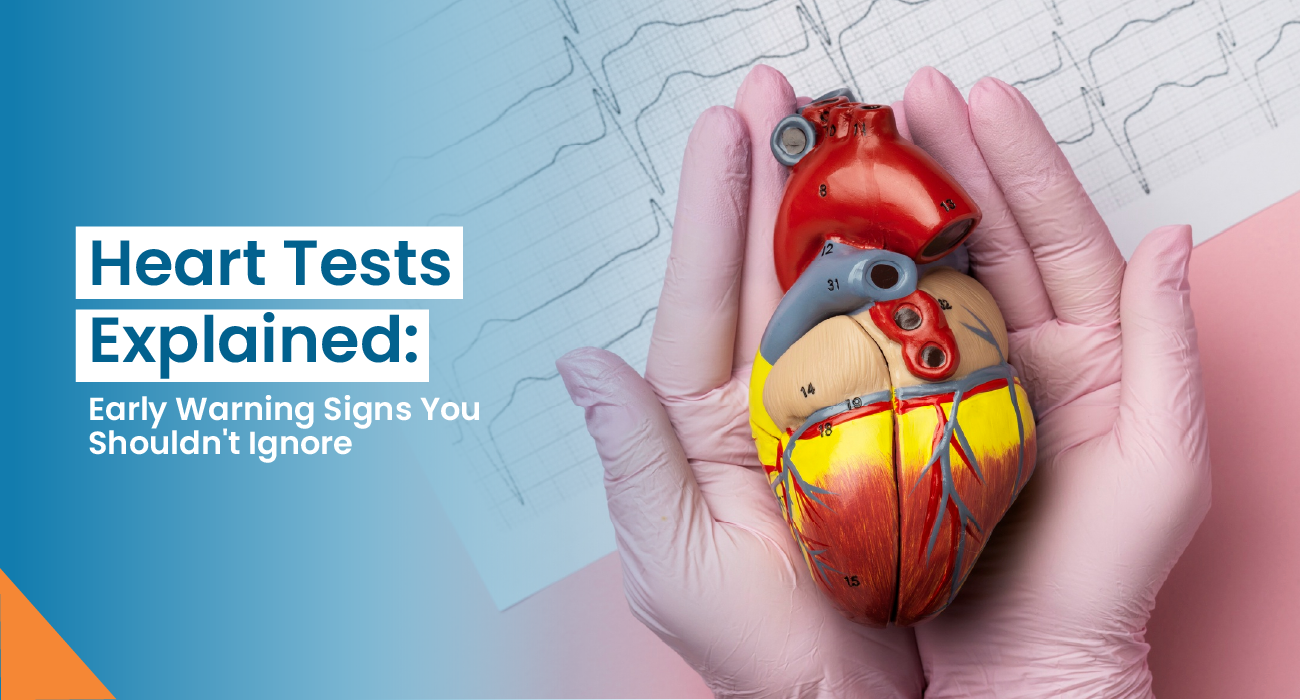
Heart Tests Explained: ECG, 2D Echo, TMT, and Coronary Angiography
09/17/2026
If your cardiologist in Jaipur has advised you to undergo any of these heart tests, such as an ECG test, 2D echo, TMT test, or coronary angiography, it’s natural to wonder: Why do I need this test? And what do these tests actually show?
These tests provide your doctor information about different aspects of your heart. For example:
- An echocardiogram checks your heart’s structure and function.
- An electrocardiogram (ECG or EKG) is a quick test to check the heartbeat.
- A TMT checks how your heart responds to physical stress.
- Coronary angiography looks for blockages in your coronary arteries.
So, one heart test is not necessarily “better” than another. Your doctor may recommend one or more of these tests depending on your condition, symptoms, medical history, risk factors, and what your doctor needs to find out.
Quick Overview: Main Heart Tests
Explore the table below to have a quick understanding of the ECG test, 2D echo, TMT test, and coronary angiography.
| Heart test | What it mainly checks | Common purpose |
| ECG test | Electrical activity and heart rhythm | Detects arrhythmias, signs of heart attack, and abnormal heart rates |
| 2D echo | Heart structure, valves, and pumping function | Evaluates heart function, valve disease, and structural problems |
| TMT test | Heart response during exercise | Looks for exercise-related changes suggesting reduced blood supply |
| Coronary angiography | Coronary arteries and blood flow | Identifies the location and severity of artery blockages |
Let’s understand each test one by one.
ECG or EKG Test
An electrocardiogram (abbreviated as EKG or ECG) is one of the most common heart tests. It is a simple, safe and painless test that records the electrical signals of the heart, including its rate and rhythm.
An ECG is a quick test to check the heartbeat. Test results can help diagnose heart attacks and irregular heartbeats (known as arrhythmias).
An ECG can help identify:
- Irregular heart rhythms or arrhythmias
- An abnormally fast or slow heart rate
- Signs of a current or previous heart attack
- Certain changes associated with reduced blood supply to the heart
- Some clues about changes in heart structure
During the test, up to 12 sticky patches called electrodes are placed on the chest and sometimes on the arms and legs. These electrodes detect electrical activity, which is displayed as waves on a screen or printed on paper.
Can ECG detect a bad heart?
Yes.
ECG can show many types of heart problems (arrhythmias, a previous heart attack, causes of chest pain, etc.), but it does not catch every issue.
2D Echo
An echocardiogram (also known as an echo or heart ultrasound) is a test that checks your heart’s structure and function.
A 2D echo is different from an ECG, which primarily examines electrical activity.
An echocardiogram uses high-frequency sound waves to create moving images of the heart as it beats. This test can help your cardiologist diagnose and treat various heart conditions.
An echocardiogram can help diagnose:
- Blood clots
- Congenital heart disease
- Cardiomyopathy
- Heart tumors
- Infective endocarditis
- Pericarditis
- Thoracic aortic aneurysm
- Valve disease
A 2D echo can provide information about:
- Heart chambers
- Heart muscle movement
- Pumping function
- Heart valves
- Blood flow through the heart
- Some structural abnormalities
- Certain congenital heart problems
Is 2D echo radioactive?
No.
A 2D echo does not use radiation.
TMT Test
A TMT test (treadmill test), also known as an exercise stress test.
An exercise stress test helps evaluate how well your heart responds during times when it's working its hardest.
A TMT may help doctors determine the following:
- Exercise-related symptoms
- Possible reduced blood supply to the heart
- Exercise capacity
- Abnormal heart rhythms triggered by exertion
How the heart responds to increasing workload
A TMT test generally involves walking on a treadmill or pedalling on a stationary bike while hooked up to an EKG to monitor your heart’s activity.
Can a TMT detect a heart blockage?
Yes, but in an indirect manner.
A TMT cannot directly show where a coronary artery is blocked. But it may show signs suggesting that the heart muscle is not getting sufficient blood during exercise.
Coronary Angiography
A coronary angiogram is a test that uses X-rays and contrast dye to visualise blood flow through the coronary (heart) arteries.
The test can show blockages in these arteries or whether they’re narrowed, usually due to atherosclerosis (hardening of the arteries caused by plaque gradually building up inside them).
A cardiologist inserts a thin catheter via an artery, commonly from the wrist or groin, and guides it toward the heart. Contrast dye is then injected while X-ray images are taken.
This can show:
- Whether a coronary artery is narrowed or blocked
- Where the blockage is located
- How severe the narrowing is
- How blood is moving through the coronary arteries
What happens if an angiogram shows a blockage?
If a significant blockage is identified, based on the individual situation, treatment options may include the following:
- Medicines
- Angioplasty and stenting,
- Bypass surgery
Conclusion
If you are experiencing the following symptoms, contact a cardiologist in Jaipur:
- Chest pain or pressure
- Breathlessness
- Unexplained fatigue
- Dizziness or fainting
- Fast or irregular heartbeat
- Reduced exercise tolerance
- A history of heart disease or significant risk factors
They can assess your symptoms and medical history and decide whether an ECG, 2D echo, TMT, coronary angiography, or another investigation is appropriate. You might feel a bit anxious before medical tests, but just remember, you’re getting valuable information to help take good care of your heart.
ECG vs 2D Echo vs TMT vs Coronary Angiography
If you are confused about which test does what, this comparison makes it simpler:
| Feature | ECG | 2D Echo | TMT | Coronary Angiography |
| Main focus | Electrical activity | Structure & function | Response to exercise | Coronary arteries |
| Exercise involved | No | Usually no | Yes | No |
| Invasive | No | No, for a standard TTE | No | Yes |
| Uses radiation | No | No | No | Yes |
| Can it assess rhythm? | Yes | Limited | Yes, during exercise | Monitoring is done, but not its main purpose |
| Can it assess pumping function? | Limited clues | Yes | Indirectly | Can provide other cardiac information but not its primary purpose |
| Can directly show coronary blockage? | No | No | No | Yes |
Disclaimer: This blog is for informational use only. For accurate information regarding heart tests, consult your doctor.