


What Happens During Brain Surgery? A Step-by-Step Breakdown
07/17/2025
In modern medicine, one of the most challenging and life-threatening procedures is brain surgery, often known as neurosurgery. Brain surgery involves a number of carefully thought-out procedures, ranging from the removal of a tumour to the treatment of a brain aneurysm, seizure control, clot drainage, or trauma management. Modern technology, accurate surgery, and careful patient care are required at every stage, from diagnosis to recuperation. Patients and their families can feel less anxious about this life-saving procedure if they understand how brain surgery operates.
Stage 1: Pre-Operative Planning
Identification and Assessment
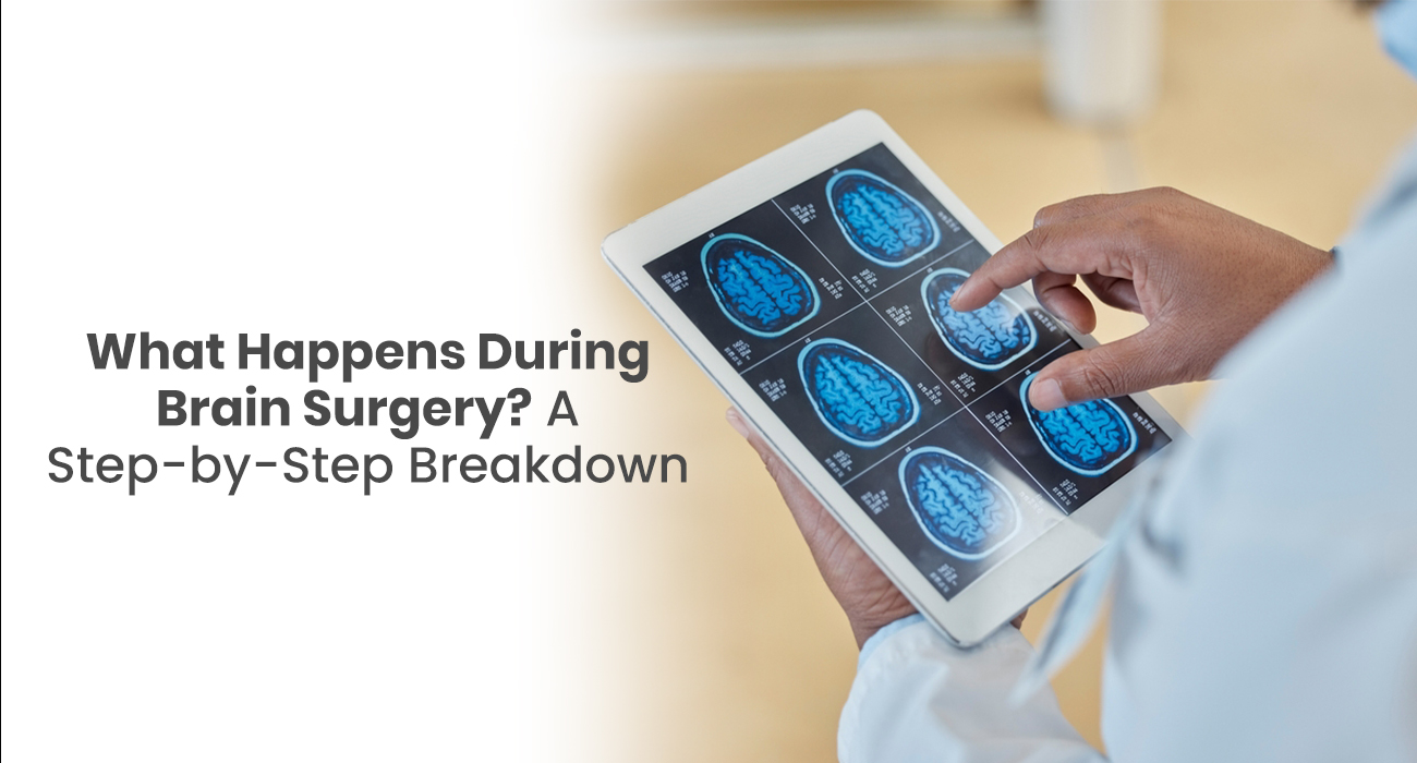
Before surgery is ever considered, a comprehensive diagnostic procedure must be completed. This includes imaging tests such as cerebral angiograms, CT scans, and MRIs. These provide neurosurgeons with a map of the precise location, size, and nature of the brain problem. To evaluate speech, motor function, memory, and vision, additional neurological testing is carried out. EEGs are occasionally used to track seizure activity. These assessments help determine whether the patient is a good candidate for surgery and guide the decision-making process.
Patient Preparation and Informed Consent
The patient and their family sit with the surgical team once the procedure is arranged. At this stage, the team shares the diagnosis, the reasons for the recommendation of surgery, and the expected outcomes. All potential dangers are fully explained, including bleeding, infection, and temporary neurological impairments. After that, patients are asked to sign an informed consent form.
Blood-thinning drugs are usually discontinued at this juncture, and pre-anesthesia assessment is performed. Patients are also instructed not to eat or drink for a number of hours prior to the procedure.
Surgical Planning and Mapping
Contemporary brain surgeries depend on accurate planning. Surgeons utilize neuronavigation systems—similar to GPS for the brain—to navigate through the procedure. Functional mapping is used to locate critical areas of the brain controlling speech, movement, and sensation, so they can be spared during the operation. The surgical team can also have pre-operative planning sessions with oncologists, radiologists, and anesthesiologists to agree on the best and safest method.
Read Also: Recovering From Brain Surgery: Cope with the situation with these tips
Stage 2: Operating Room Setup
Anesthesia and Positioning
The patient is brought into the operating room and given general anaesthesia for the procedure. A neuro-anesthesiologist keeps a close eye on the patient's vital indicators, such as blood pressure, heart rate, and oxygen saturation. A specialised head holder is used to place and immobilise the head. This design keeps the brain steady throughout the treatment.
Setting Up a Sterile Field
The surgical site is cleansed thoroughly once the patient has been positioned and put to sleep. The skin is sterilised and the scalp in the area that will be operated on is shaved. After that, sterile drapes are placed over the patient, creating an absolutely sterile operating room to prevent infection.
Read Also: Neurosurgery for Brain Tumors: Diagnosis, Treatment Options, and Prognosis
Stage 3: Craniotomy
Making the Skin Incision
A tailored scalp incision is performed depending on the area of interest in the brain. The incision may be camouflaged behind the hairline. The skin and tissues are softly retracted, exposing the skull below.
Removing the Bone Flap
The neurosurgeon makes tiny holes—burr holes—in the skull using a high-speed drill. These are linked by a special instrument known as a craniotome to cut away a section of skull called a bone flap. This flap is carefully removed and will be replaced once surgery is completed.
Accessing the Brain
Under the skull is the dura mater, a thick protective membrane of the brain. The dura mater is opened by the surgeon with caution to reveal the brain tissue. Careful solutions are employed to ensure the moistening of the brain and avoid damage. Mild retractors might be inserted gently to slide brain tissue aside so the surgeon can have unobstructed sight into the operated area.
Read Also: Understanding Brain Tumor Symptoms and Treatment Options
Stage 4: Accessing and Treating the Lesion
Microsurgical Exposure
With the use of an operating microscope and fine instruments, the surgeon starts to lay open the area of interest. Whether tumor, aneurysm, hematoma, or vascular malformation, the aim is to access the lesion with as minimal disturbance of healthy brain tissue as possible. Neuronavigation techniques are frequently employed to assist in distinguishing between diseased and normal tissue.
Tumor Removal
For tumors of the brain, the surgeon will initially decompress by debulking or incising the tumor. Next, the margins of the tumor are dissected away from adjacent brain structures. Some procedures employ fluorescence-guided approaches, where dyes illuminate tumor cells in real time to guarantee a total extirpation.
Aneurysm Clipping
If the patient has an aneurysm of the brain, the surgeon exposes the involved blood vessel. A titanium clip of small size is inserted into the neck of the aneurysm to prevent blood flow into the weakened portion and to avoid rupture. The positioning of the clip is verified through intraoperative angiography or Doppler monitoring.
Hematoma Evacuation or AVM Resection
In cases of bleeding or arteriovenous malformations (AVMs), the procedure involves suctioning out blood clots or removing abnormal blood vessels. This must be done very carefully to avoid damage to nearby healthy brain regions.
Stage 5: Intraoperative Monitoring
Brain Monitoring for Safety
During the procedure, advanced monitoring devices monitor the brain and nerve function. They use sensory and motor evoked potentials to monitor changes in nerve function. During surgeries close to speech centers, patients are sometimes under anesthesia for half of the procedure—this is called an awake craniotomy—avoiding any damage to critical brain functions such as language.
Managing Bleeding and Brain Safety
Bleeding during the operation is controlled through the use of a combination of bipolar cautery (a heat-sealing device for blood vessels), hemostatic agents, and constant irrigation. This makes it possible to obtain an unobstructed view and minimize complications.
Stage 6: Closure of the Skull and Scalp
Dura Repair
After the lesion has been removed or treated, the dura mater is checked by the surgeon. If necessary, it is covered by a patch using a graft to obtain a watertight closure. This prevents cerebrospinal fluid (CSF) leakage after surgery.
Replacement of the Bone Flap
The bone flap that has been previously excised is then fixed back in position using titanium plates or bioresorbable clamps. This reconstructs the skull and shields the brain.
Closure of the Scalp
Lastly, the soft tissues and skin are closed in layers. For the deeper tissues, internal sutures are employed; for the skin, staples or sutures are utilized. A sterile dressing is placed to guard against infection.
Stage 7: Immediate Post‑Operative Recovery
ICU monitoring
The patient is taken to the Intensive Care Unit (ICU) or high-dependency unit post-operatively. Nurses and physicians continually monitor neurological status, which consists of consciousness, pupil response, speech, and extremity movement. Pain relief and antibiotics or anti-seizure medication are given as needed.
Post-Operative Imaging
A CT scan is typically done within 24 hours of surgery. This allows the team to ensure that there is no bleeding, swelling, or other complications within the body. If all is well, the patient can be transferred to a general hospital room in one or two days.
Stage 8: Rehabilitation and Physical Recovery
Physical Therapy
When the patient is stable, physical rehabilitation starts. Physical therapists assist patients in the recovery of movement, balance, and independence. Early mobilization will prevent the formation of blood clots and accelerate recovery.
Speech and Cognitive Therapy
For procedures close to brain areas involved with speech or memory, patients might need individualized cognitive and language therapy. These therapies restore lost function and develop compensatory strategies if deficits persist.
Wound Care and Observation
The surgical area is monitored closely for infections, such as fever, redness, or drainage. Stitches or staples are normally removed at 7–10 days.
Stage 9: Discharge Planning and Long-Term Recovery
Instructions for Home
Instructions given on discharge include specific details regarding medications, activity restrictions, and wound care. Heavy lifting is prohibited, as is vigorous activity or bending, for several weeks. Explicit follow-up appointment schedules are set with the neurosurgeon or oncologist.
Continued Imaging
Follow-up imaging—typically MRI or CT scans—should be done one month after operation and as needed thereafter. These assist with monitoring tumor regrowth or verifying aneurysm clip or other repair stability.
Mental and Emotional Support
Emotionally, brain surgery can also affect a person. The patient can develop mood swings, anxiety, or even a change in personality. Psychological guidance is usually suggested to guide both the patient and caregivers in dealing with the process.
Stage 10: Returning to Normal Life
Resuming Activities
The majority of patients recover slowly and resume work, exercise, and daily activities in 4 to 12 weeks. The duration varies based on the nature of the surgery and the recovery of the patient. For patients who have high-demand occupations or need mental accuracy, neuropsychological testing can be performed prior to returning to work.
Surveillance and Prevention
Some conditions, like brain tumors, may require additional treatment such as radiation or chemotherapy. Others, like aneurysms, may require periodic follow-up angiograms to ensure long-term success. Preventive care—including managing blood pressure and lifestyle changes—is key to reducing recurrence risk.
Conclusion
Brain surgery is the most remarkable achievement of contemporary medicine. While the thought of having surgery on the brain may be frightening, knowing what is done at each step—preoperatively, intraoperatively, and postoperatively—will diminish considerable fear and anxiety. With sophisticated equipment, expert neurosurgeons, and well-organized recovery programs, results improve annually.